
Ultrasound Podcast
About
Available on
Community
93 episodes

I am so happy to share with you our latest video on how to perform an ultrasound guided pericardiocentesis! This video has gone through a few revisions but I think this one is my best one so far (although I do concede that I may be a little bit biased🙃) this video already assumes that you know how to tell a difference between a pericardial effusion and pericardial tamponade. If you need a review, please check out the YouTube/website for a refresher! Hope you enjoy! 🌐 Connect with Us 🌐 If you want more education goodness, check out the links below! Check out our longitudinal, year-long and virtual ultrasound fellowship here: https://ultrasoundleadershipacademy.com Check out our FREE content on our website: https://www.coreultrasound.com https://www.coreultrasound.com/ Check out our courses here: https://courses.coreultrasound.com Check out our question bank here: https://courses.coreultrasound.com/collections/q-bank By reading the content of this website, you agree not to use this information as medical advice to treat any medical condition in either yourself or others, including but not limited to patients that you are treating. Consult your own physician for any medical issues that you may be having. This entire disclaimer applies to any and all content produced by Jailyn Avila, Ben Smith or Core Ultrasound. Under no circumstances shall any of the contributors on this content be responsible for damages arising from use of this (or other) content. This content should not be used in any legal capacity whatsoever, including but not limited to establishing “standard of care” in a legal sense or as a basis for expert witness testimony. No guarantee is given regarding the accuracy of any statements or opinions made on the website. The content of this website is formed by our own opinions and do not represent the views or opinions.

This week on the podcast we have a new version of the 5 Minute Sono video on the sonographic evaluation of the IVC. The IVC is not going to primarily diagnose anything (usually!), but instead is used to evaluate the central venous pressure (CVP). If the IVC is plump and not collapsing, it means a high CVP, and if it is small and collapsing a lot, it means low CVP. This is most helpful when evaluating a patient in shock, as it can help push you towards or away from specific diagnoses (such as septic shock vs cardiogenic shock). This is actually the third revision of this educational content! First one was in 2015 and the second revision was in 2021. I’m humbled to have been creating medical education content for as long as I have and I try and keep my videos fresh and up to date :) Hope you enjoy! 🌐 Connect with Us 🌐 If you want more education goodness, check out the links below! Check out our longitudinal, year-long and virtual ultrasound fellowship here: https://ultrasoundleadershipacademy.com Check out our FREE content on our website: https://www.coreultrasound.com https://www.coreultrasound.com/ Check out our courses here: https://courses.coreultrasound.com Check out our question bank here: https://courses.coreultrasound.com/collections/q-bank By reading the content of this website, you agree not to use this information as medical advice to treat any medical condition in either yourself or others, including but not limited to patients that you are treating. Consult your own physician for any medical issues that you may be having. This entire disclaimer applies to any and all content produced by Jailyn Avila, Ben Smith or Core Ultrasound. Under no circumstances shall any of the contributors on this content be responsible for damages arising from use of this (or other) content. This content should not be used in any legal capacity whatsoever, including but not limited to establishing “standard of care” in a legal sense or as a basis for expert witness testimony. No guarantee is given regarding the accuracy of any statements or opinions made on the website. The content of this website is formed by our own opinions and do not represent the views or opinions.

In this week’s video, we’re showing you our second episode of the new Ultrasound Leadership Academy Image Review: From Motion to Meaning. The Ultrasound Leadership Academy is a non-profit whose sole function is to educate medical providers on best practices while incorporating ultrasound into their daily practice. We offer many different tiers of support, including dedicated image review, personalized curriculum and one-on-one virtual hangouts. Click the link to learn more! 🌐 Connect with Us 🌐 Check out our FREE content on our website: https://www.coreultrasound.com Check out our courses here: https://courses.coreultrasound.com Check out our question bank here: https://courses.coreultrasound.com/collections/q-bank Check out our in-person courses here: https://www.soundandsurf.com/

In the last episode I showed you part one of the interview I had with Dr. Terren Trott discussing the velocity time integral and how it can be used to help manage our sick patients. In this episode, I share with you the thrilling conclusion! If you want to learn this technique AND MORE, check out our upcoming in-person mini-course SONOLAB, March 5th in sunny San Diego. (Click here for more info! https://www.soundandsurf.com/the-sono-lab/p/san-diego-2024) 🌐 Connect with Us 🌐 If you want more education goodness, check out the links below! Check out our longitudinal, year-long and virtual ultrasound fellowship here: https://ultrasoundleadershipacademy.com Check out our FREE content on our website: https://www.coreultrasound.com https://www.coreultrasound.com/ Check out our courses here: https://courses.coreultrasound.com Check out our question bank here: https://courses.coreultrasound.com/collections/q-bank

Have you ever wondered how much fluid is too much in your critical ill patients? Join us in this enlightening session with Dr. Terren Trott MD, an emergency medicine AND critical physician who also completed an ultrasound fellowship, as we talk about how the VTI can be used in assessing volume responsiveness in our critically ill patients. This interview is going to be split up into TWO parts. Stay tuned for part 2 coming soon! If you want to learn this technique AND MORE, check out our upcoming in-person mini-course SONOLAB, March 5th in sunny San Diego. (Click here for more info! https://www.soundandsurf.com/the-sono-lab/p/san-diego-2024) And lastly, in case you’d like to check out the article we talked about at the beginning, here’s the link: https://pubmed.ncbi.nlm.nih.gov/30997543/ 🌐 Connect with Us 🌐 If you want more education goodness, check out the links below! Check out our longitudinal, year-long and virtual ultrasound fellowship here: https://ultrasoundleadershipacademy.com Check out our FREE content on our website: https://www.coreultrasound.com https://www.coreultrasound.com/ Check out our courses here: https://courses.coreultrasound.com Check out our question bank here: https://courses.coreultrasound.com/collections/q-bank

In this episode, Michael Macias and I chat about some barriers and benefits of using ultrasound in community (non or less academic) emergency departments. Any place where medicine is practiced should be held to similar standards, and POCUS should be considered standard of care. We discuss a study done at our home institution where we looked at a quite large sample of ultrasounds and various providers to give insight on which exams are common and some potential missed opportunities! Check out the podcast for more info! References: __ __ 🌐 Connect with Us 🌐 If you want more education goodness, check out the links below! Check out our longitudinal, year-long and virtual ultrasound fellowship here: https://ultrasoundleadershipacademy.com Check out our FREE content on our website: https://www.coreultrasound.com https://www.coreultrasound.com/ Check out our courses here: https://courses.coreultrasound.com Check out our question bank here: https://courses.coreultrasound.com/collections/q-bank

In part 2 of this amazing interview, Dr. Trott and I discuss specifics on ultrasound and how it can benefit critical care and emergency medicine physicians. We discuss our favorite position for central venous catheters (Spoiler alert: We both love right subclavians the most!) and other interesting insights. Check it out! 🌐 Connect with Us 🌐 If you want more education goodness, check out the links below! Check out our longitudinal, year-long and virtual ultrasound fellowship here: https://ultrasoundleadershipacademy.com Check out our FREE content on our website: https://www.coreultrasound.com https://www.coreultrasound.com/ Check out our courses here: https://courses.coreultrasound.com Check out our question bank here: https://courses.coreultrasound.com/collections/q-bank

Have you ever wanted to ask an intensive care doc about how they utilize POCUS in their daily practice? What about a critical care doc who is also emergency medicine and POCUS-fellowship trained? I got to sit down (in person!) with Dr. Terren Trott, who is a California transplant in Kentucky who has some amazing insights to share with us. Check it out! 🌐 Connect with Us 🌐 If you want more education goodness, check out the links below! Check out our longitudinal, year-long and virtual ultrasound fellowship here: https://academy.ultrasoundleadershipacademy.com Check out our FREE content on our website: https://www.coreultrasound.com https://www.coreultrasound.com/ Check out our courses here: https://courses.coreultrasound.com Check out our question bank here: https://courses.coreultrasound.com/collections/q-bank

In this episode, I walk you through basic machine operation for the Sonosite Xporte, including selecting the appropriate probe/setting, inputting patient (and your) information, how to record and how to export to review later or store in your institution (please check with your institution on appropriate technique for storing images/clips!) In the future, we will be publishing more content like this, that should be helpful to all comers. Please leave a comment if there’s a specific machine you’d like me to work on next! 🌐 Connect with Us 🌐 If you want more education goodness, check out the links below! Check out our longitudinal, year-long and virtual ultrasound fellowship here: https://academy.ultrasoundleadershipacademy.com Check out our FREE content on our website: https://www.coreultrasound.com https://www.coreultrasound.com/ Check out our courses here: https://courses.coreultrasound.com Check out our question bank here: https://courses.coreultrasound.com/collections/q-bank

I am so happy to share with you all part two of my first interview with Dr. Michael Macias! In this podcast we talk about a single topic: fracture management. Please enjoy this second part of my interview with Dr. Macias and we can't wait to hear from you and learn how you all utilize ultrasound in the management of suspected or confirmed fractures Oh, and if you want to learn how to identify fractures on ultrasound and how to do hematoma blocks, check out our newest 5 Minute Sono on the topic: https://coreultrasound.com/fracture-and-hematoma-blocks/ 🌐 Connect with Us 🌐 If you want more education goodness, check out the links below! Check out our longitudinal, year-long and virtual ultrasound fellowship here: https://academy.ultrasoundleadershipacademy.com Check out our FREE content on our website: https://www.coreultrasound.com https://www.coreultrasound.com/ Check out our courses here: https://courses.coreultrasound.com Check out our question bank here: https://courses.coreultrasound.com/collections/q-bank

I'm so happy to finally be able to sit down with Dr. Michael Macias and discuss a few things that I've been meaning to ask him. Mike graduated from an ultrasound fellowship and then begin working as a community-based physician where he became the Ultrasound Director for Emergent Medical Associates, as well as the UHS Southern California Residency Consortium, he is the Assistant Emergency Department Director AND is the founder of the POCUS Atlas. It was impossible to fit in everything that I wanted to talk with him about in one episode! Please enjoy this first part of my interview with Dr. Macias where we discuss: How to use ultrasound in a community based-setting to place transvenous pacers, how to use right heart ultrasound effectively in both confirmed and suspected PE as well as fracture management. 🌐 Connect with Us 🌐 If you want more education goodness, check out the links below! Check out our longitudinal, year-long and virtual ultrasound fellowship here: https://academy.ultrasoundleadershipacademy.com Check out our FREE content on our website: https://www.coreultrasound.com Check out our courses here: https://courses.coreultrasound.com Check out our question bank here: https://courses.coreultrasound.com/collections/q-bank

The Ultrasound Leadership Academy is a non-profit that was founded 10 years ago the the same people who first started the Ultrasound Podcast (Mike Malin, Matt Dawson and Mike Stone), and our mission is to spread the highest-quality ultrasound education to all. This video is a new series to highlight some of the features of ULA, including dedicated ultrasound image/clip review, focused on ways to improve adequate image acquisition, as well as personalized online video-conference one-on-one mentoring. Check out https://academy.ultrasoundleadershipacademy.com learn more, and please feel free to reach out to admin@ultrasoundleadershipacademy.com! This episode includes intrauterine device (IUD) identification the FAST exam, renal calyceal rupture“ 🌐 CONNECT WITH US 🌐 If you want more education goodness, check out the links below! Check out our longitudinal, year-long and virtual ultrasound fellowship here: https://academy.ultrasoundleadershipacademy.com Check out our FREE content on our website: https://www.coreultrasound.com https://www.coreultrasound.com/ Check out our courses here: https://courses.coreultrasound.com Check out our question bank here: https://courses.coreultrasound.com/collections/q-bank

Welcome to our latest video where we explore the crucial role of point-of-care ultrasound in early detection of ectopic pregnancies. 🌐 In just a few minutes, we guide you through the process of using ultrasound to identify signs and symptoms, ensuring timely and effective medical intervention. 🤰 What's Inside? Discover the key features of an ectopic pregnancy ultrasound, understand how to differentiate it from a normal intrauterine pregnancy, and empower yourself with the knowledge to enhance patient care. 👍 Don't Forget to Like, Share, and Subscribe! If you found this video informative, give it a thumbs up, share it with your colleagues, and subscribe to Core Ultrasound for more insightful content. 🔔 Turn on Notifications: Hit the notification bell so you never miss an update from us! 🔗 Learn More: Interested in incorporating point-of-care ultrasound into your medical practice? Find resources and training opportunities in the links below. If you want more education goodness, check out the links below! Check out our longitudinal, year-long and virtual ultrasound fellowship here: https://academy.ultrasoundleadershipacademy.com Check out our FREE content on our website: https://www.coreultrasound.com Check out our courses here: https://courses.coreultrasound.com Check out our question bank here: https://courses.coreultrasound.com/collections/q-bank

Welcome to our latest video, where we delve into using point-of-care ultrasound for determining the fetal heart rate using M-mode. 🔍 What's Inside? In this short and informative video, we guide you through the steps of using M-mode on a point-of-care ultrasound machine to accurately assess the fetal heart rate. Whether you're a healthcare professional or an ultrasound enthusiast, this tutorial breaks down the process in a clear and concise manner. The main point: Using M-mode instead of pulsed-wave Doppler likely involves less energy transmitted to the fetus. 👍 Don't Forget to Like, Share, and Subscribe! If you found this video informative, give it a thumbs up, share it with your colleagues, and subscribe to Core Ultrasound for more insightful content. 🔔 Turn on Notifications: Hit the notification bell so you never miss an update from us! 🔗 Learn More: Interested in incorporating point-of-care ultrasound into your medical practice? Find resources and training opportunities in the links below. If you want more education goodness, check out the links below! Check out our longitudinal, year-long and virtual ultrasound fellowship here: https://academy.ultrasoundleadershipacademy.com Check out our FREE content on our website: https://www.coreultrasound.com Check out our courses here: https://courses.coreultrasound.com Check out our question bank here: https://courses.coreultrasound.com/collections/q-bank

In this 5 Minute Sono video we are going to discuss the basics on how to identify an intrauterine pregnancy using your bedside ultrasound machine. Follow our step-by-step guide to using point-of-care ultrasound for intrauterine pregnancy identification. From preparation to scanning techniques, we cover it all, including how to interpret key indicators on the ultrasound screen. 👍 Don't Forget to Like, Share, and Subscribe! If you found this video informative, give it a thumbs up, share it with your colleagues, and subscribe to Core Ultrasound for more insightful content. 🔗 Learn More: Interested in incorporating point-of-care ultrasound into your medical practice? Find resources and training opportunities in the links below. If you want more education goodness, check out the links below! Check out our longitudinal, year-long and virtual ultrasound fellowship here: https://academy.ultrasoundleadershipacademy.com Check out our FREE content on our website: https://www.coreultrasound.com https://www.coreultrasound.com/ Check out our courses here: https://courses.coreultrasound.com Check out our question bank here: https://courses.coreultrasound.com/collections/q-bank

In this 5 Minute Sono video we are going to discuss some basic scanning techniques involving OB/Ob Gyn. This is a must watch video for point of care providers wanting to learn the basics on how to evaluate pregnant patients and patients with suspected uterine and ovarian pathology! Stay tuned for upcoming videos with specific pregnancies, including ectopic, pregnancies, how to assess by ability and the fetal heart rate! 🌐 CONNECT WITH US 🌐 If you want more education goodness, check out the links below! Check out our longitudinal, year-long and virtual ultrasound fellowship here: https://academy.ultrasoundleadershipacademy.com Check out our FREE content on our website: https://www.coreultrasound.com https://www.coreultrasound.com/ Check out our courses here: https://courses.coreultrasound.com Check out our question bank here: https://courses.coreultrasound.com/collections/q-bank
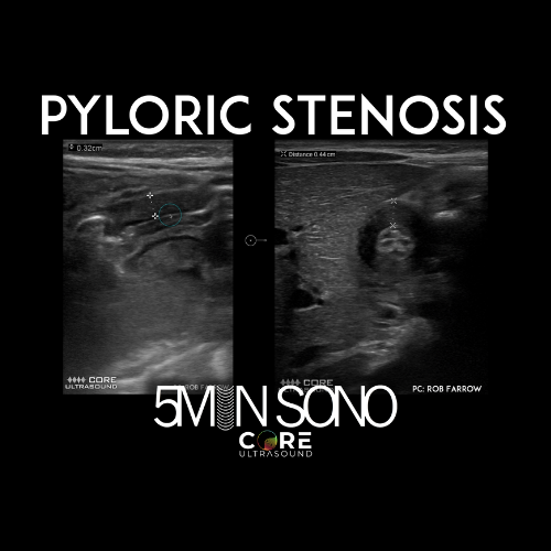
Pyloric stenosis is a relatively common condition in infants that can lead to severe vomiting, dehydration, and weight loss. Timely and accurate diagnosis is crucial for effective treatment. Bedside ultrasound has become a valuable tool for diagnosing pyloric stenosis quickly and non-invasively. Learn how to effectively perform the exam definitely within the reach of the non-radiologist clinician. Check out the video for more details! 🌐 CONNECT WITH US 🌐 If you want more education goodness, check out the links below! Check out our longitudinal, year-long and virtual ultrasound fellowship here: https://academy.ultrasoundleadershipacademy.com Check out our FREE content on our website: https://www.coreultrasound.com Check out our courses here: https://courses.coreultrasound.com Check out our question bank here: https://courses.coreultrasound.com/collections/q-bank

In this video, we will guide you through the sonographic evaluation of a suspected ankle effusion using bedside ultrasound. We will also demonstrate how to accurately perform an ankle aspiration and injection. Check out this latest 5 Minute Sono video for details on how to do it! 🌐 CONNECT WITH US 🌐 Check out our BRAND-NEW course: SonoAnatomy here: https://courses.coreultrasound.com/courses/sonoanatomy Check out our longitudinal, year-long and virtual ultrasound fellowship here: https://academy.ultrasoundleadershipacademy.com Check out our FREE content on our website: https://www.coreultrasound.com https://www.coreultrasound.com/ Check out our courses here: https://courses.coreultrasound.com Check out our question bank here: https://courses.coreultrasound.com/collections/q-bank
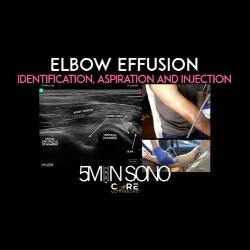
In this Five Minute Sono video, Jailyn discusses how to identify an elbow effusion, and then how to aspirate fluid or inject medications. This is a potentially very difficult procedure made easier by bedside ultrasound. Check out the video to learn more! 🌐 CONNECT WITH US 🌐 Check out our BRAND-NEW course: SonoAnatomy here: https://courses.coreultrasound.com/courses/sonoanatomy Check out our longitudinal, year-long and virtual ultrasound fellowship here: https://academy.ultrasoundleadershipacademy.com Check out our FREE content on our website: https://www.coreultrasound.com Check out our courses here: https://courses.coreultrasound.com Check out our question bank here: https://courses.coreultrasound.com/collections/q-bank
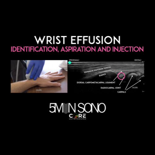
Ultrasound has high accuracy for diagnosing a wrist effusion. As a bonus, it ALSO increases the accuracy of aspirations and injections. Check out the latest 5 Minute Sono video for details on how to do it! 🌐 CONNECT WITH US 🌐 Check out our BRAND-NEW course: SonoAnatomy here: https://courses.coreultrasound.com/courses/sonoanatomy Check out our longitudinal, year-long and virtual ultrasound fellowship here: https://academy.ultrasoundleadershipacademy.com Check out our FREE content on our website: https://www.coreultrasound.com Check out our courses here: https://courses.coreultrasound.com Check out our question bank here: https://courses.coreultrasound.com/collections/q-ban
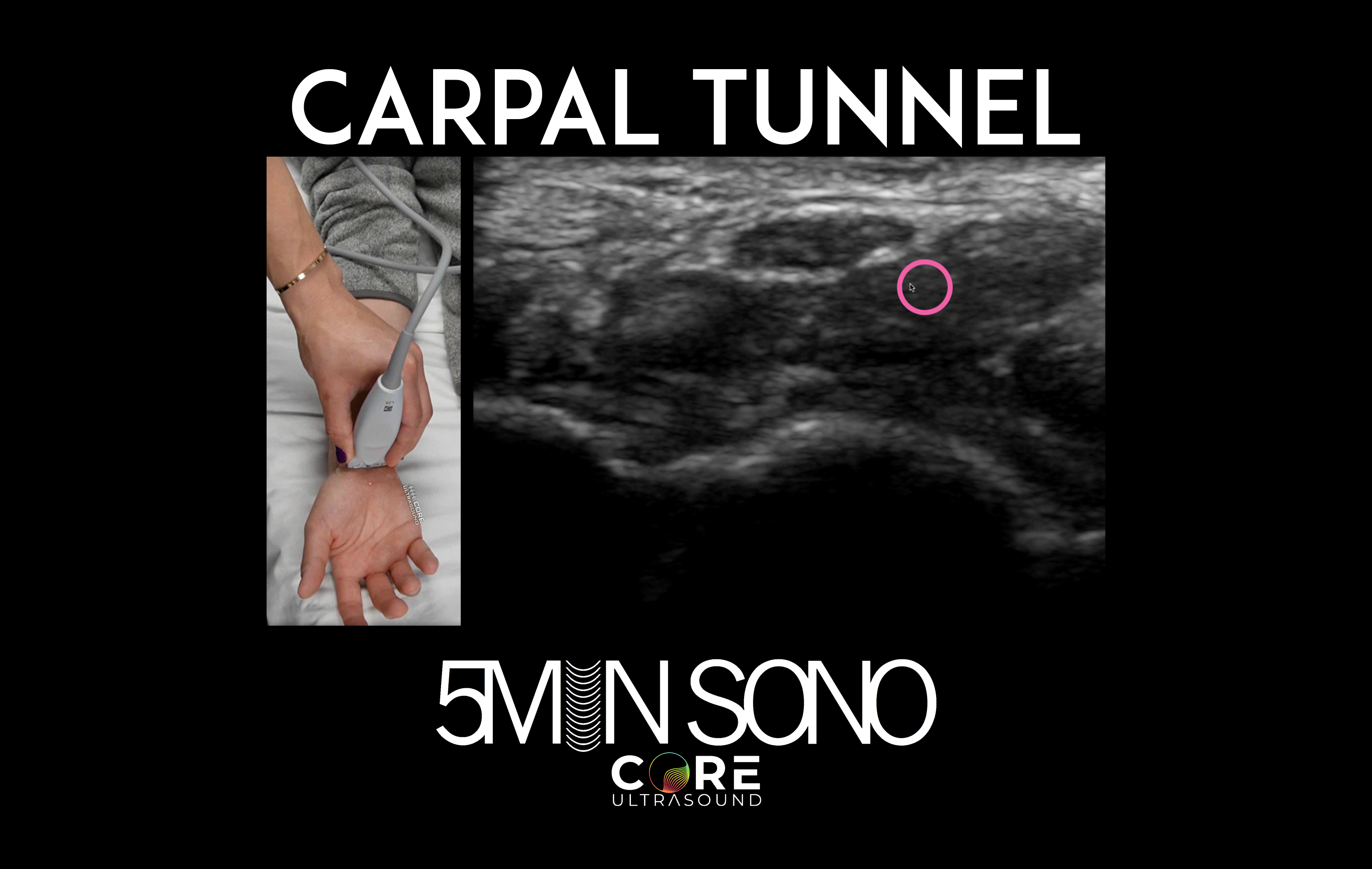
Carpal tunnel syndrome affects millions of people and may be tricky to definitely diagnose. Bedside ultrasound can be a phenomenal adjunct in the evaluation in patients suspected of having this debilitating illness. Check out our 5 Minute Sono video to learn how to accurately diagnose carpal tunnel using a simple measurement of the median nerve inside the carpal tunnel! 🌐 CONNECT WITH US 🌐 If you want more education goodness, check out the links below! Check out our longitudinal, year-long and virtual ultrasound fellowship here: https://academy.ultrasoundleadershipacademy.com Check out our FREE content on our website: https://www.coreultrasound.com Check out our courses here: https://courses.coreultrasound.com Check out our question bank here: https://courses.coreultrasound.com/collections/q-bank

🔍 Unlock the secrets of ultrasound with our concise guide on Pulsed Wave and Continuous Wave Doppler, and how they can revolutionize your medical practice. 🎓🌐 Discover the key differences, applications, and advantages of each method in this brief, 5 Minute Sono video. 🌐 CONNECT WITH US 🌐 If you want more education goodness, check out the links below! Check out our longitudinal, year-long and virtual ultrasound fellowship here: https://academy.ultrasoundleadershipacademy.com Check out our FREE content on our website: https://www.coreultrasound.com Check out our courses here: https://courses.coreultrasound.com Check out our question bank here: https://courses.coreultrasound.com/collections/q-bank
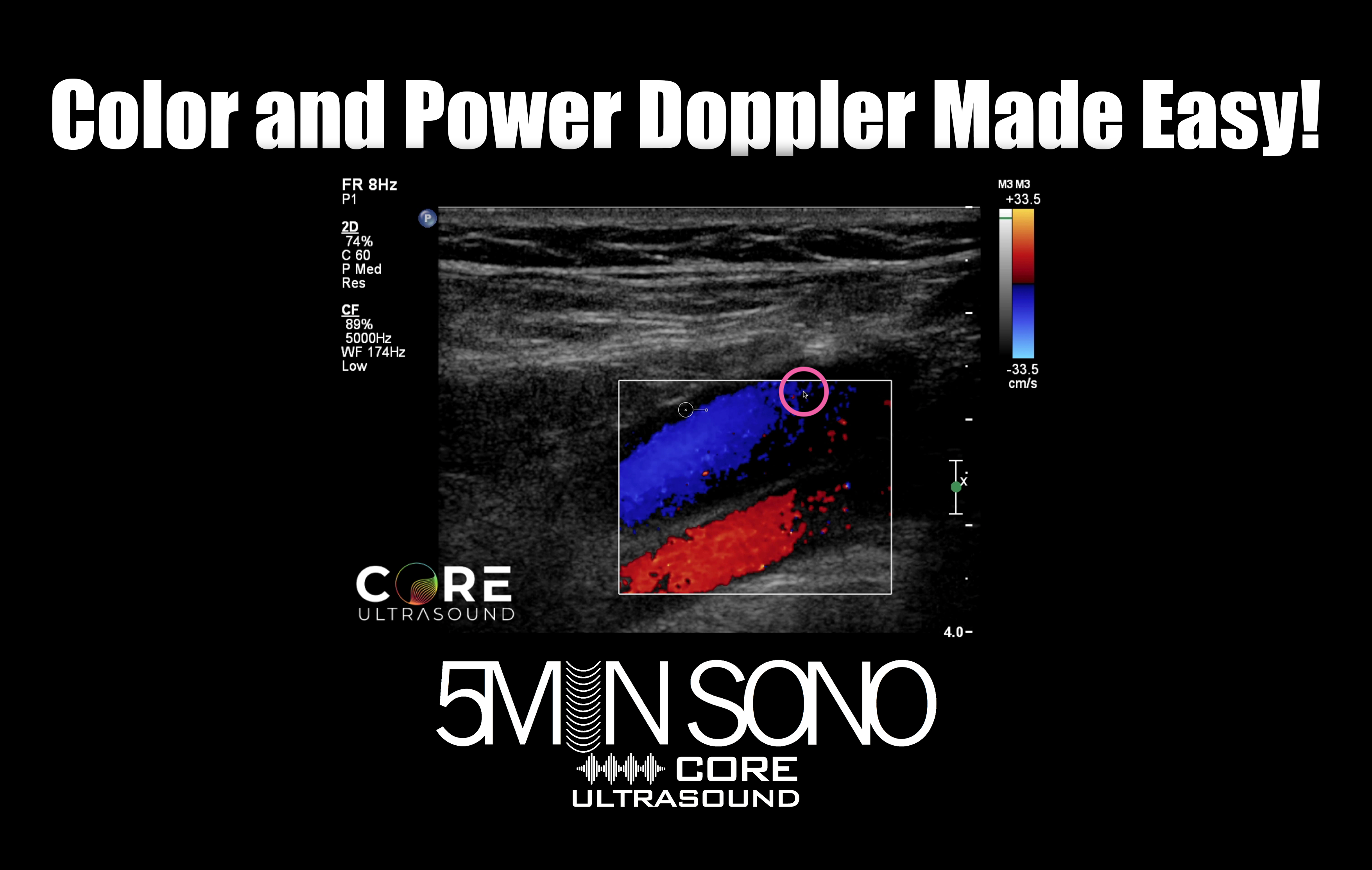
🌈 UNRAVELING THE MAGIC OF COLOR DOPPLER 🌈 Welcome to Core Ultrasound! In this 5 Minute Sono educational video, we dive into the fascinating world of ultrasound imaging and explore the principles behind Color and Power Doppler. Color Doppler is a crucial technique used in medical ultrasound to visualize blood flow within the body. By assigning colors to moving blood cells, we can easily identify the direction and speed of blood flow in real-time. But how does it all work? Join us as we break down the basics behind color mapping and demonstrate its clinical significance through captivating examples. 🧠 EDUCATIONAL AND INFORMATIVE 🧠 Whether you're a medical professional, student, or simply curious about medical imaging technologies, this video is designed to be informative and accessible to all. Our expert explanation and insightful visuals will take you on a journey through the inner workings of these Doppler techniques, helping you gain a deeper understanding of their applications and limitations. 🔔 DON'T FORGET TO SUBSCRIBE! 🔔 If you found this video helpful or interesting, make sure to hit the "Subscribe" button and click the notification bell so you won't miss any of our future uploads. We have a lot more exciting content coming your way! 🌐 CONNECT WITH US 🌐 If you want more education goodness, check out the links below! Check out our longitudinal, year-long and virtual ultrasound fellowship here: https://academy.ultrasoundleadershipacademy.com Check out our FREE content on our website: https://www.coreultrasound.com Check out our courses here: https://courses.coreultrasound.com Check out our question bank here: https://courses.coreultrasound.com/collections/q-bank
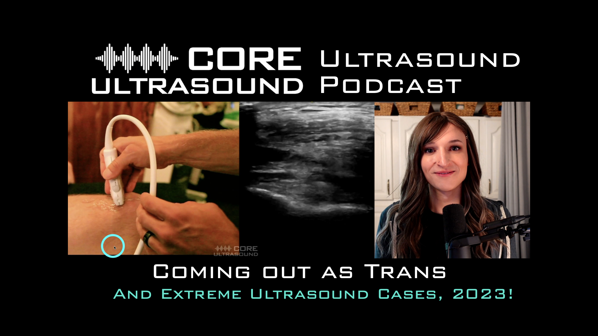
I’m so proud to be living my truth and sharing my story with you all! Join me in this special episode where I share just a little bit of my journey of coming out to the rest of the world as a transgender woman. Also, there are 5 other reasons to watch: I’ll share with you my favorite ultrasound cases of the past few months. I hope you enjoy this wide-ranging talk on some pretty important and amazing topics! Check out our FREE content on our website: https://www.coreultrasound.com Check out our courses here: https://courses.coreultrasound.com Check out our question bank here: https://courses.coreultrasound.com/collections/q-bank
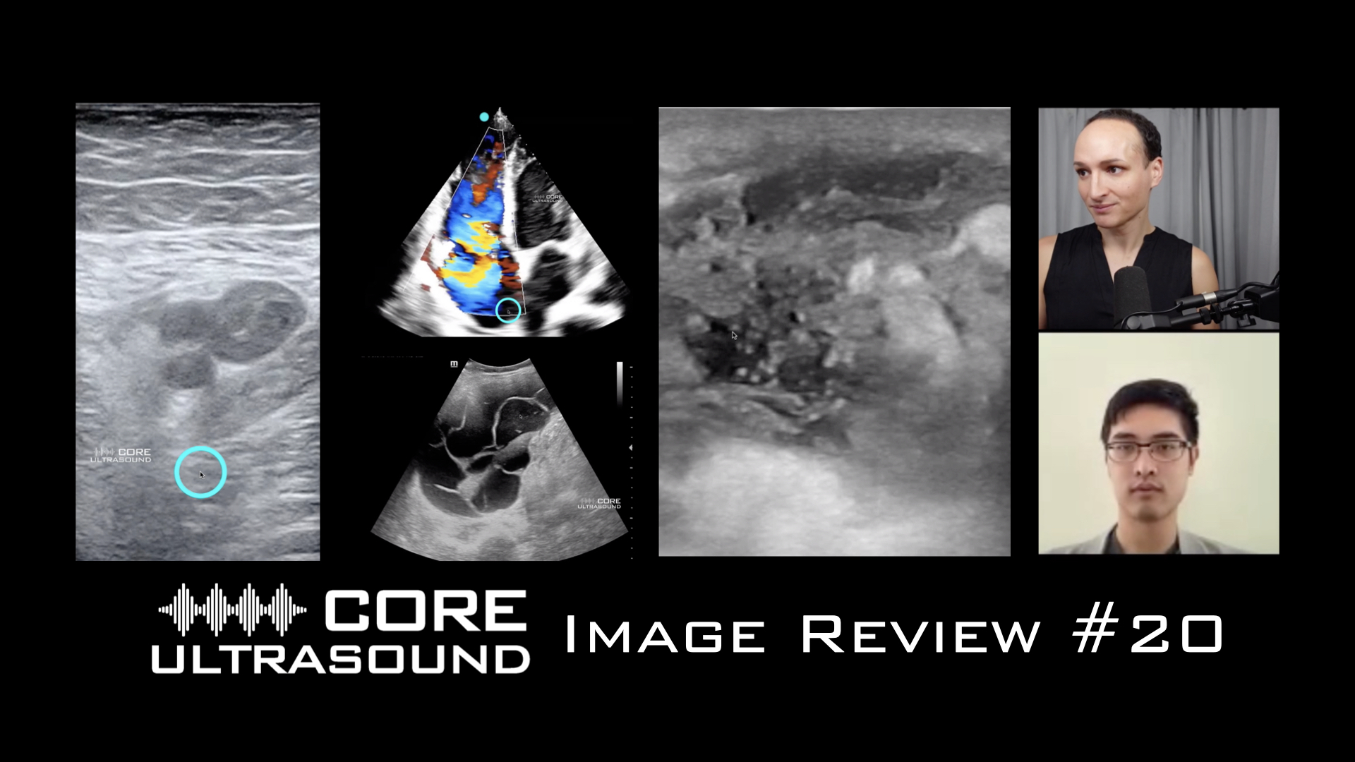
At long last, we are finally revitalizing the image review segment of our channel and website! The purpose of image review sessions is to just go through a hodgepodge of ultrasound clips seen at various places that I (or guests to the show) have worked. The idea first came up when I was doing image reviews of your sessions when I was the Ultrasound Director at the University of Kentucky. There was so much good information and knowledge translation that I felt like I needed to be shared with a broader audience than with my residents, fellows and faculty. Sometimes we have specific themes and sometimes it’s just cool ultrasounds that we’ve collected. We try to give one or more learning tips per ultrasound clip. If you have any questions on this specific one, please leave the question in the comment! If you have any clips that you would like to submit, please email! Check out our longitudinal, year-long and virtual ultrasound fellowship here: https://academy.ultrasoundleadershipacademy.com Check out our FREE content on our website: https://www.coreultrasound.com Check out our courses here: https://courses.coreultrasound.com Check out our question bank here: https://courses.coreultrasound.com/collections/q-bank
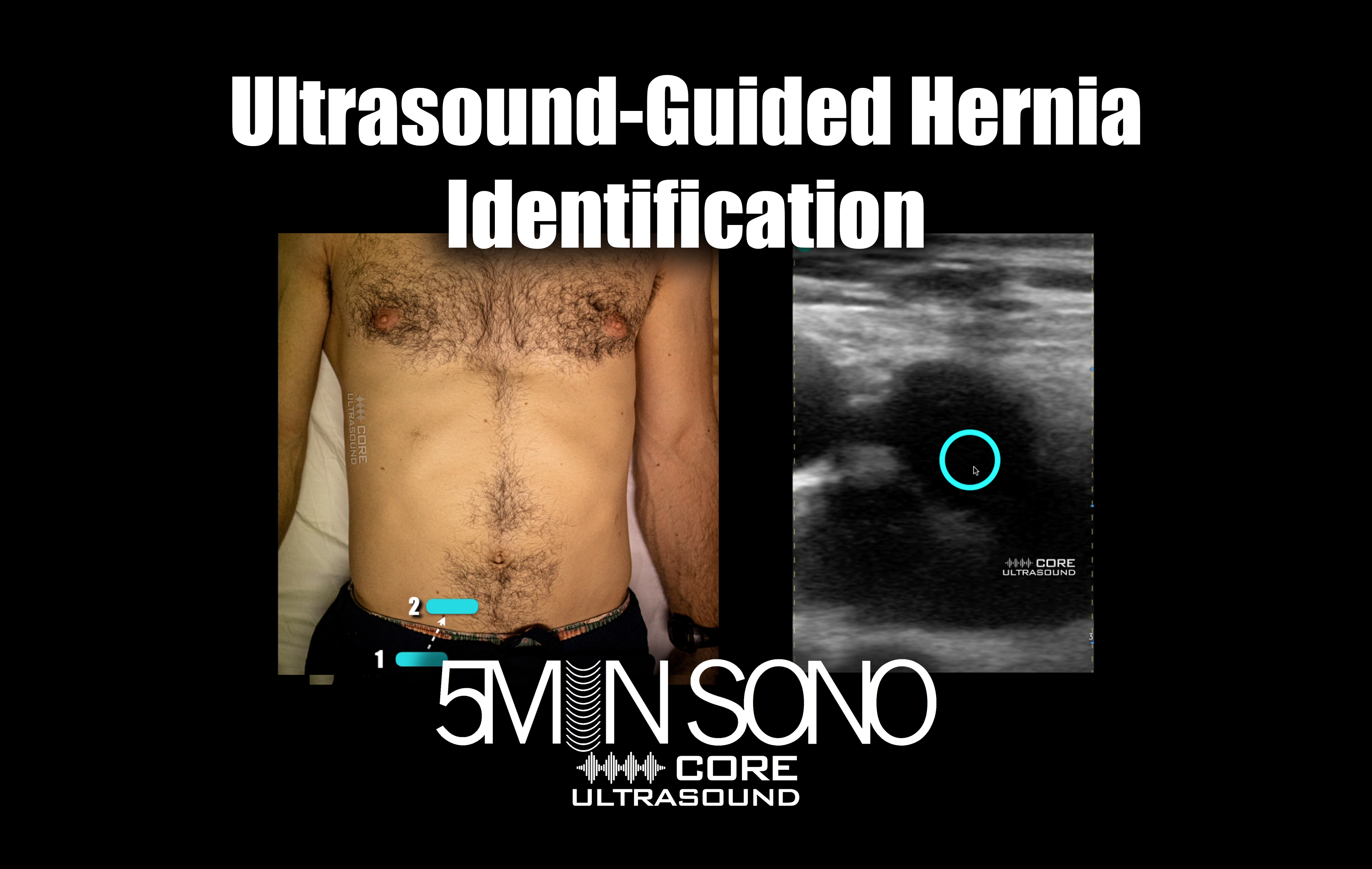
In this 5 Minute Sono video, we'll guide you through the process of using ultrasound imaging to identify and diagnose inguinal and femoral hernias. Hernias can be difficult to identify with physical examination alone (and may actually be missed on CT!), but with ultrasound imaging, healthcare professionals can detect hernias at the bedside with greater accuracy and confidence. This video will walk you through the steps of performing an ultrasound scan for hernia identification, including patient positioning, ultrasound probe placement, and image interpretation. You'll learn about the different types of hernias and how they appear on ultrasound images, as well as the key features to look for when assessing for a hernia. Check out our FREE content on our website: https://www.coreultrasound.com Check out our courses here: https://courses.coreultrasound.com Check out our question bank here: https://courses.coreultrasound.com/collections/q-bank
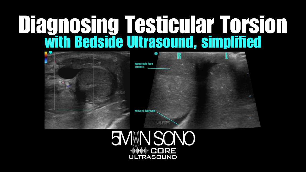
In this Five Minute Sono video, Paul Khalil (with the help of Rob Farrow), walks us through the bedside diagnosis of testicular torsion. This disease is quite important to diagnose as early as possible and using this tool may dramatically increase the chance of testicular salvage. Check out our FREE content on our website: https://www.coreultrasound.com Check out our courses here: https://courses.coreultrasound.com Check out our question bank here: https://courses.coreultrasound.com/collections/q-bank
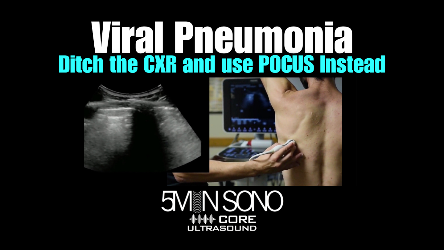
In this Five Minute Sono I show you how to diagnose a viral pneumonia using bedside ultrasound. While the most common application these days is probably for COVID-19 pneumonia, these techniques also can be used to diagnose any viral pulmonary infections. Bacterial pneumonia has a slightly different appearance, and I’ll post that video later in the week! Check out our FREE content on our website: https://www.coreultrasound.com Check out our courses here: https://courses.coreultrasound.com Check out our question bank here: https://courses.coreultrasound.com/collections/q-bank
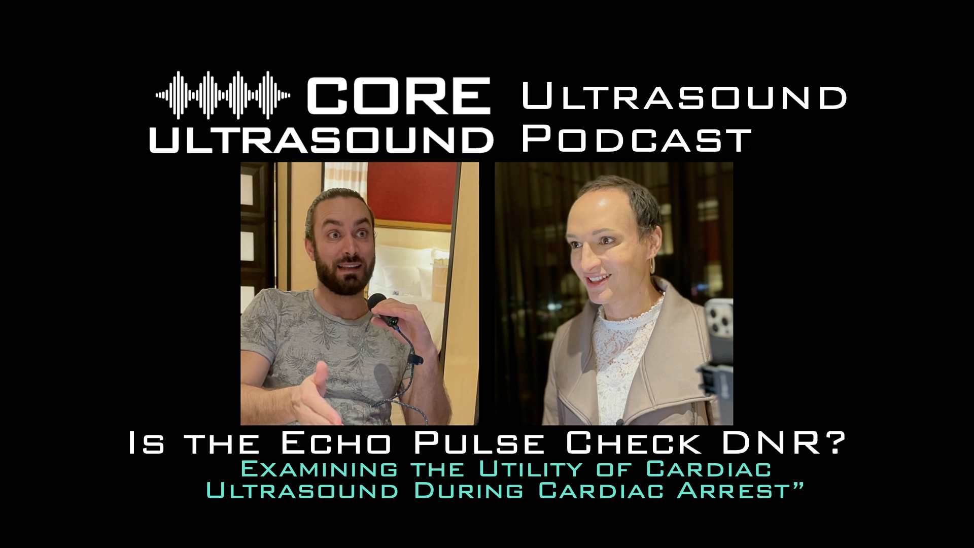
Cardiac POCUS can be dramatically useful and life-saving in cardiac arrest. I like to think about it in three different broad categories: Procedural, identifying reversible causes, and guiding therapy. This video isn’t a discussion regarding its utility for procedures or diagnosing reversible causes, but it is talking about how maybe the echo pulse check is not the best way to assess for adequate perfusion during pulse checks. Terren Trott, MD and I discuss our thoughts on two articles demonstrating how carotid and femoral artery ultrasound can be utilized during pulse checks. Check out the publications we will be discussing on Pubmed: PMIDs 35131404 and 35792305. Check out our FREE content on our website: https://www.coreultrasound.com Check out our courses here: https://courses.coreultrasound.com Check out our question bank here: https://courses.coreultrasound.com/collections/q-bank
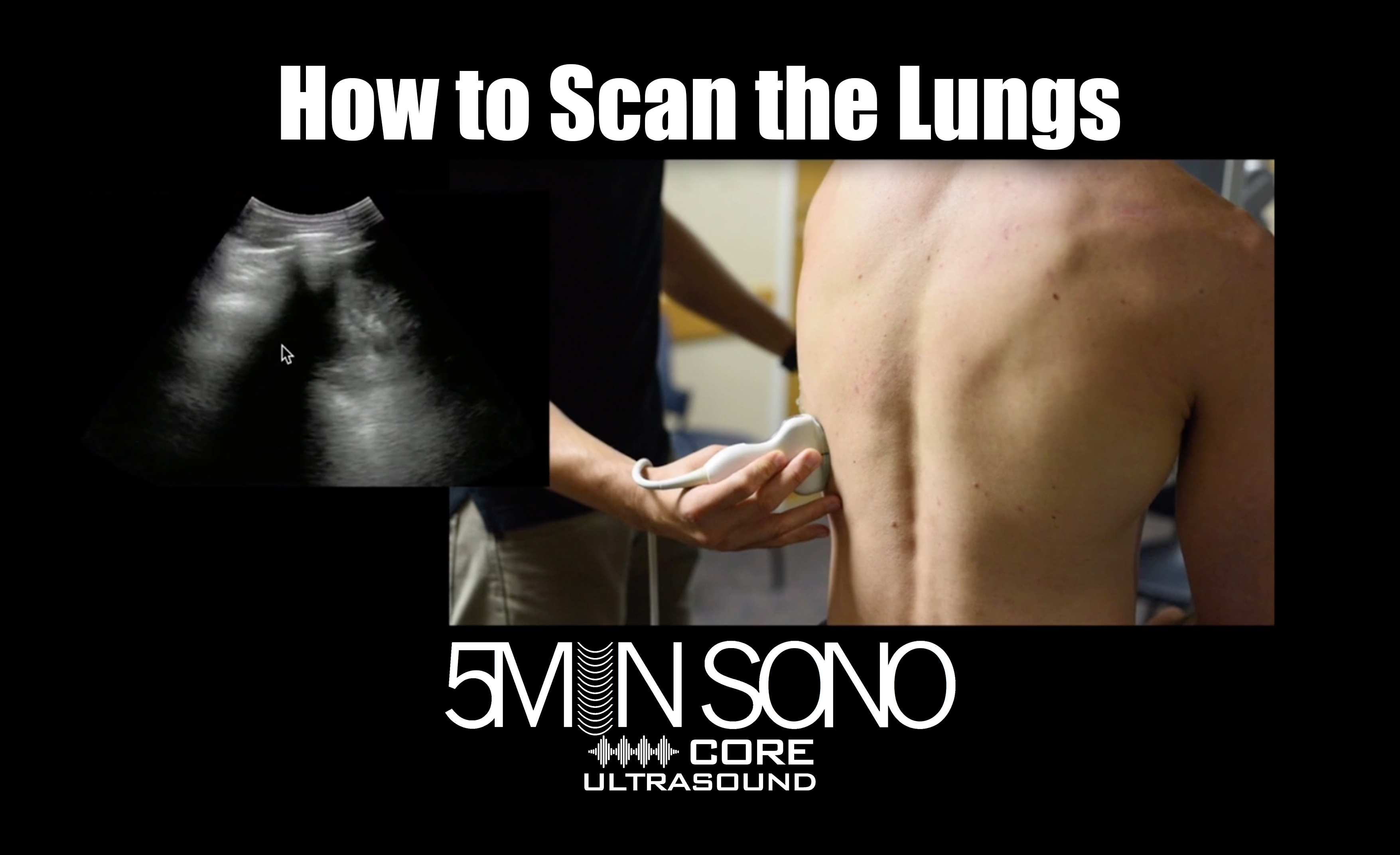
In this five minute sono video, I’m going to walk you through how to get started scanning the lungs. The most common applications for performing an ultrasound of the lungs include pulmonary edema (traditionally seen in heart failure exacerbations but can be present in plenty of other pathologies), pneumothorax and pleural effusion. Check out our FREE content on our website: https://www.coreultrasound.com Check out our courses here: https://courses.coreultrasound.com Check out our question bank here: https://courses.coreultrasound.com/collections/q-bank